Electrical Properties of Tissue
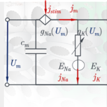
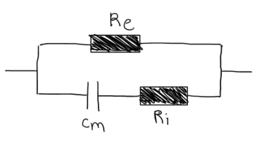
Biological tissue behaves capacitively in an alternating electric field and exhibits frequency-dependent changes. Thus, it can be represented qualitatively as a circuit of two resistors and a capacitor or constant phase element. Here, the resistors Re and Ri represent the extracellular and intracellular space, respectively, and the capacitance Cm represents the blocking behavior of the cell membrane. This means that the current flows around the cells at low frequencies (path via Re ) and can only penetrate the cell membrane above a certain threshold value.
At first glance, the electrical impedance can be determined quite easily by measuring the current flow when the tissue is excited with AC voltage. However, many influencing factors such as surface pressure, temperature or the arrangement of the measuring electrodes play a role - and so does the condition of the tissue. Already at the beginning of the last century it could be determined experimentally that tumor tissue has a different impedance compared to healthy tissue. Structural changes such as the compression of the ECM and the enlargement of the cell nucleus, as well as the altered physiology, are responsible for this.
Impedance Measurements
The aim of the experiments is to generate frequency spectra covering the relevant ranges of the so-called alpha and beta dispersion. The sensor used plays a decisive role in the quality of the data obtained. To minimize unwanted polarization effects and to achieve the highest possible precision, a four-wire measurement is essential. This requires a total of four electrical contact points with the tissue - two to conduct the signal and two to measure the applied voltage. The arrangement of these electrodes on the available surface also has an influence, especially with regard to the application case of bladder cancer, where the endoscopic access is max. 2mm. While with conventional four-wire measurements the electrodes are traditionally arranged in a row, the minimally invasive version offers a staggered or even concentric arrangement.
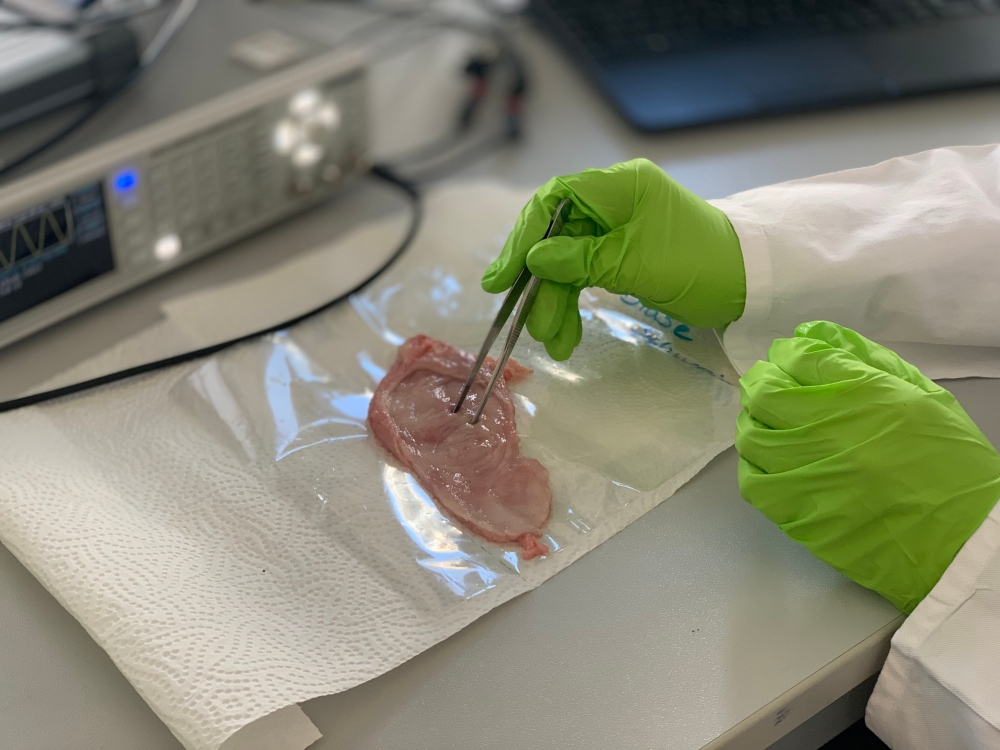
The first tests and the comparison of different measurement arrangements are performed on pig bladders and serve to optimize the sensor before it is then tested on human ex-vivo samples.
Classification
In the case of breast cancer, approaches already exist for classifying healthy and malignant tissue using neural networks or linear discriminant analysis. In the case of colorectal cancer, even a differentiation of the different tumor stages has been shown based on measurements of relative permittivity. Project A5 will investigate both model-based approaches and data-based approaches to classification.

Zoltan Lovasz
M.Sc.PhD Student A5
- Profile page
- +49 711 685 66300
- Write e-mail
- Raum 1.39

Carina Veil
Dr.-Ing.
Oliver Sawodny
Prof. Dr.-Ing. habil. Dr. h.c.Sprecher des GRK 2543