Fringe Projection Using 3D-Printed Micro-Optics for Model-Based Tissue Differentiation Based on Optical and Elastic Properties
The primary goal of all new interventional procedures in surgery is to combine minimal invasiveness and high effectiveness with short treatment duration and low complication rates. A central aspect in oncology is the differentiation between malignant target structures and the surrounding tissue during surgery. Frozen section diagnostics is considered the gold standard of intraoperative tissue differentiation. During this procedure, the tissue is taken to a laboratory immediately after resection, cut in layers, stained, and examined histopathologically. The surgery usually has to be interrupted until the result is available. In addition, therapy (tumor resection) and diagnosis (histological examination) are separated both spatially and temporally.
The fusion of novel, multimodal sensor systems using machine learning methods yields a high potential that exceeds that of the individual sensors. The multimodal sensor systems are intended to support surgeons in making decisions between resection and tissue preservation as a supplement to frozen section diagnostics. Under this multimodal approach, sensor systems are to be developed and used. Due to the changed tissue characteristics, this should enable a fast and intraoperative assessment of the tissue.
In order to enable greatest possible functional preservation for the patient, a small-volume identification of the tumor boundaries is necessary. The central topic of research project A1 is the discrimination of tissue boundaries based on elastic and optical parameters using optical methods. This approach is based on the fact that cancerous tissue has a different morphology as well as proliferation and therefore metabolism than healthy tissue.
The increased growth of tumor tissue leads to a denser but less organized structure of the malignant tissue and to changes in the extracellular matrix (ECM). The increased density of the ECM leads to changes in the elastic parameters of the tissue. This serves as an approach to tissue differentiation, as the elasticity is quantifiable.

Furthermore, biological tissue can be regarded as an optically diffuse, turbid medium, since scattering usually dominates over absorption when light propagates through the medium. Similarly to elastic parameters, these optical parameters are also quantifiable. Based on chromophore concentrations in tissue, absorption and scattering coefficients can be determined metrologically. Tumor tissue contains different concentrations of these chromophores than healthy tissue due to an altered cell metabolism and structure. This provides an additional approach to differentiate between tissue types.
Diffuse optical imaging can be performed in reflectance or transmission configuration. Examples of such measurement methods are diffuse optical tomography or spatial frequency domain imaging using structured illumination.
Scientific questions
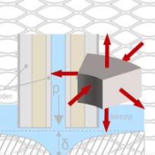
The aim of the project is the development of a multimodal and miniaturized optical sensor system, which should allow spatially resolved identification of deep tissue. In this context, already conceptually successfully tested depth-resolved optical sensor principles will be evaluated and combined regarding miniaturization (SFDI and elastographic measurement) to develop a spatially resolved differentiation of tumor margins compared to healthy tissue.
The research questions to be answered within the project is whether the combination of optical deformation and spatially resolved light scattering measurements can enable faster and improved (model-based) tissue differentiation and where its limits are. It will be investigated how these measurement methods can be combined in a multimodal probe and realized as a minimally invasive and intraoperatively applicable device.
Methods and solution approaches
SFDI will be used to differentiate deeper tissue structures based on optical properties. For this purpose, a miniaturized fringe projector, which has been already been successfully tested for elastographic measurements, will be extended to create a multimodal probe that enables tissue differentiation by combining elastographic as well as spatially resolved light scattering and absorption measurements.

The elastic parameters will also be compared and verified with theoretical simulation models. The aim of this research project is to develop a sensitive multimodal miniaturized probe for endoscopic use, which allows the acquisition of the elastic as well as optical parameters of the tissue at hand.
Editors

Ömer Atmaca
M.Sc.PhD Student A1
- Profile page
- +49 711 685 60846
- Write e-mail
- Room: 1.255B

Valese Aslani
Dr.-Ing.PhD Student A1 (Alumni)
- Profile page
- +49 711 685 60955
- Write e-mail
- Raum 1.10

Alois Herkommer
Prof. Dr. rer. nat.Manager of Subproject A1
- Profile page
- +49 711 685 69871
- Write e-mail
- Room: 1.239